
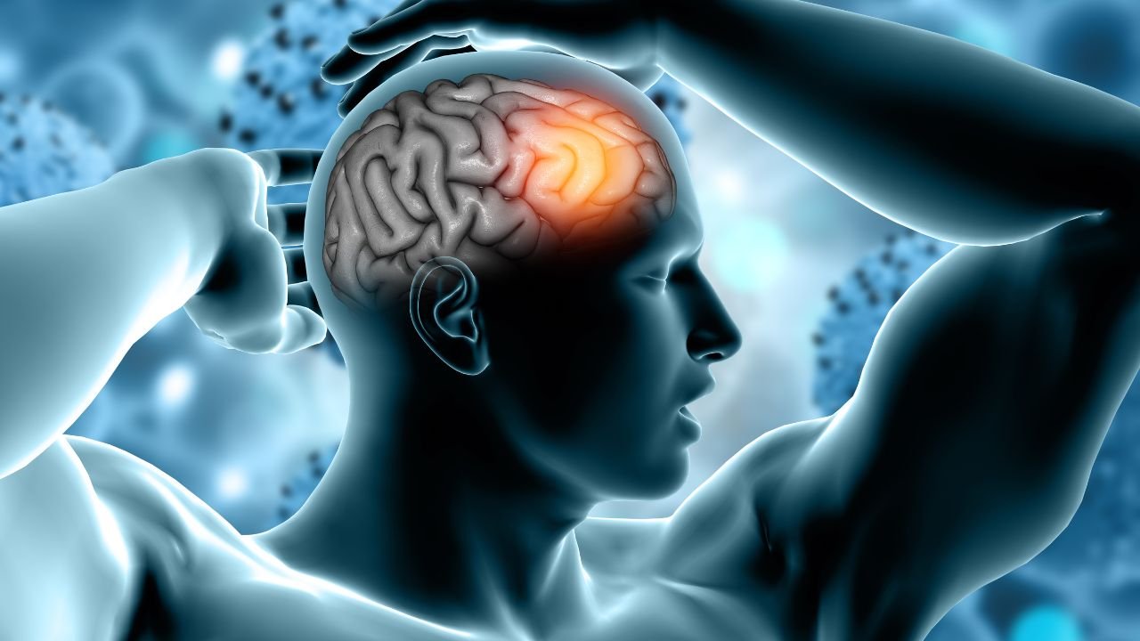
Stroke is a condition that suddenly affects a person’s ability to speak, walk, or move a part of the body. In many patients, some lost abilities return spontaneously within a few weeks of the stroke. Although very few achieve a complete recovery, most patients remain with some degree of disability. Despite this, it is interesting to note that most patients experience spontaneous recovery.
But the question is—when some brain cells are permanently destroyed during a stroke, how does a person recover? Does the brain “remap” itself and create new cells? Or do the surviving cells take over the lost functions?
To answer these questions, UCLA Health neurologists—Dr. William Zeiger and Dr. Dr. Carlos Portera-Cailliau has recently conducted research that challenges conventional wisdom.
What is a stroke and how does it affect the brain?
A stroke occurs when blood flow to a part of the brain is suddenly cut off—usually due to a blood clot. This causes cells in that area to die and the brain’s functions to be disrupted. Depending on the part of the brain affected, a person’s abilities—such as speaking, walking, recognizing objects, or feeling—are affected.
In the 19th century, French physician Paul Broca discovered that damage to a specific part of the brain’s frontal lobe could cause a person to lose speech. This discovery marked the beginning of brain “mapping”—identifying which part is responsible for which function.
To date, scientists have been striving to make this mapping more precise. However, limited technological tools have made it difficult to understand the brain’s functioning at a microscopic level.
Brain “Remapping” Theory – A New Challenge to an Old Idea
It has long been believed that the brain “remaps” itself after a stroke—that is, the damaged area is taken over by surrounding healthy areas.
Previous research in both animals and humans has shown that brain activity patterns change after a stroke. This led scientists to believe that after damage to one part of the brain, another part takes over.
But the latest study from UCLA has called this assumption into question.
New Research: Brain Cells Don’t Change Their Function
Dr. Zeiger and Dr. Portera-Cayo conducted this study on mice. They used an advanced technique called two-photon fluorescence microscopy—in which neurons (brain cells) glow when they are active, allowing scientists to see which cells are firing when.
Using this technique, scientists wanted to understand whether the neurons that survive a stroke change their function and take over for the damaged cells.
In this experiment, they targeted brain regions associated with the whiskers of mice. Mice use their whiskers to perceive their environment. Each whisker is connected to a specific neuron group. When neurons associated with a particular whisker were destroyed, they examined whether neurons associated with other whiskers could compensate.
The results were surprising—nothing of the sort happened. Neither did nearby cells take over the damaged cells’ functions, nor did “surround-responsive” neurons (those that respond slightly to stimulation from nearby whiskers) become more active. Rather, their activity decreased further.
An interesting example – understand with an office story
Dr. Portera-Cayo explained this process with a simple example. He said, “Imagine that some employees in a department’s HR office suddenly leave their jobs. Work will be affected initially, but the department will try to have other employees take over temporarily.”
This is what the brain remapping theory assumes. But this study found that this doesn’t happen in the brain—other neurons don’t shift from their original function to new ones.
This means that the lost part of the brain doesn’t automatically recover.
Spontaneous recovery: How recovery occurs remains a mystery
Although the study clarified that brain cells don’t spontaneously change their function, some patients experience improvement. The reason for this remains a mystery to science.
The researchers believe that future studies may help understand how to stimulate surviving cells to take over the functions of destroyed cells.
Dr. Zeiger says, “This study points the way. Now we must understand that if the brain cannot remap itself, we must find ways to enable cells to perform new functions.
Future Prospects: The Beginning of New Thought in Stroke Treatment
This study is not only poised to transform the current understanding of brain science, but is also extremely important for the future of stroke treatment. If scientists can identify which cells can remap the brain,If biological or chemical signals can activate brain cells, it’s possible that faster and more complete recovery after stroke may be possible in the future.
Furthermore, this research suggests that treatment after stroke shouldn’t be limited to rehabilitation. We need to develop therapies that directly affect the behavior of cells—enabling them to take over lost functions.
Conclusion: The brain’s complexity remains a mystery
This UCLA study once again highlights the complexity of the human brain. It shows that the brain is not just a “self-correcting” machine, but a highly precise and controlled system where each cell has its own specific function.
Although this research is still in its early stages and is based only on animal models, its results could have profound implications for medicine. If scientists succeed in understanding how to “train” brain cells to perform new functions in the future, a revolutionary change in the field of stroke recovery will be possible.
In short, the brain doesn’t change itself as much after a stroke as we previously thought. Improvement does occur, but the actual biological reasons behind it are still not fully understood. This new discovery from UCLA is a concrete step in that direction—one that not only provides a new perspective for science, but could also provide a new ray of hope for millions of stroke patients in the future.
